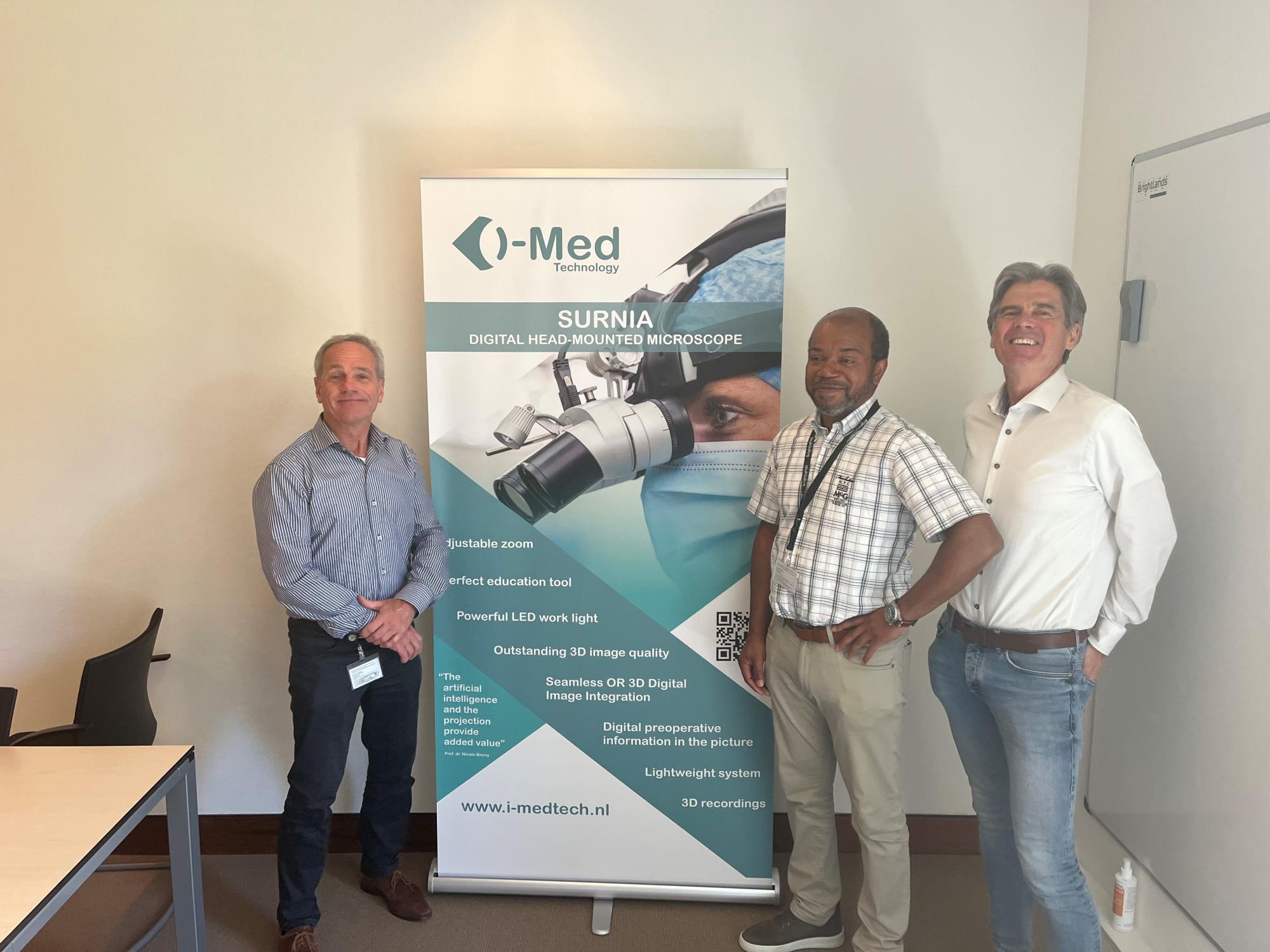
About i-Med Technology
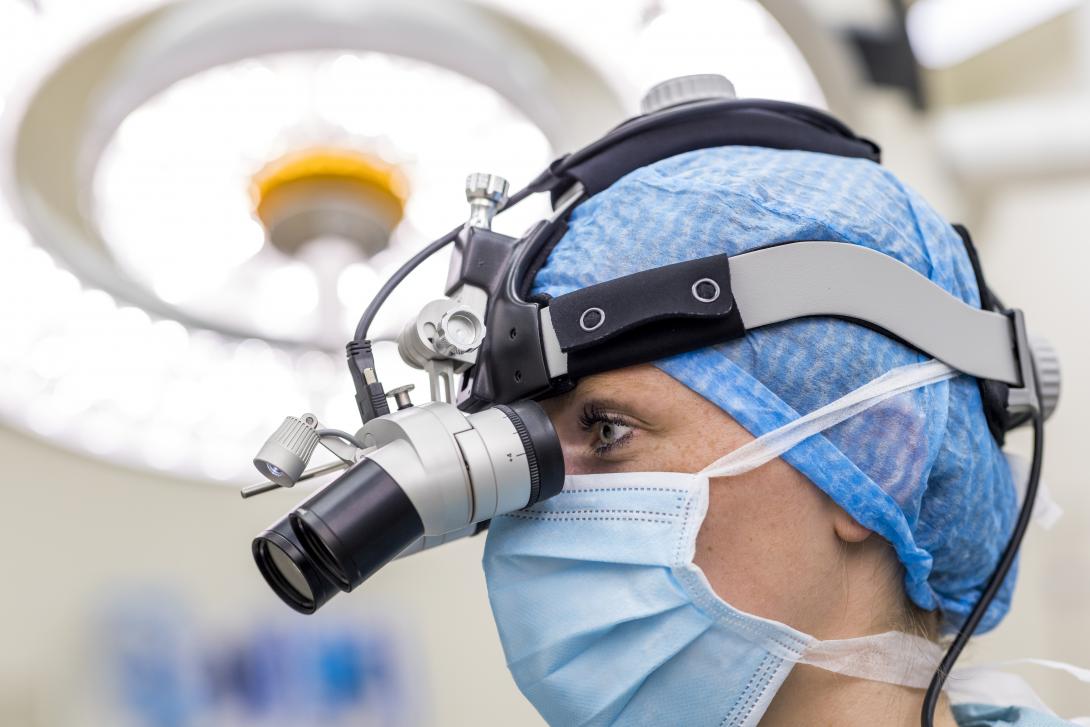
i-Med Technology, based at the Brightlands Maastricht Health Campus, has developed a successor to tools such as microscopes and magnifying glasses: A head-worn digital microscope with razor-sharp 3D image quality, zoom function and a powerful LED lamp. In the process, a camera films the operating table. The images are then digitally projected into the surgeon's eyes without delay. Bystanders inside and outside the OR can watch on screens in real time "through the surgeon's eyes," and add augmented reality layers in real time, such as MRI and CT scans. 3D recording is also possible for education and analysis.
Anyone imagining medical procedures of the future quickly thinks of (robotic) surgeons wearing VR goggles, infrared images illuminating vessels in the body and lifelike 3D recordings. That future is closer than imagined. With the Digital Head Mounted Microscope (DHM) developed by i-Med Technology for use in the operating room, surgeons have relevant information at their fingertips at a glance and can work even more accurately. "We are growing fast and are now also entering the dental market," says Jaap Heukelom, co-founder of i-Med.
Highly precise, microscopic procedures are increasingly the order of the day in the operating room. Even now, surgeons are using tools such as microscopes and magnifying glasses to properly perform these procedures. However, these tools are limited and cannot be integrated with new digital developments.
Now i-Med Technology, located at the Brightlands Maastricht Health Campus, has developed a successor to these tools: A head-worn digital microscope with razor-sharp 3D image quality, zoom function and a powerful LED lamp. A camera hereby films the operating table. The images are then digitally projected into the surgeon's eyes without delay. Bystanders inside and outside the OR can watch on screens in real time "through the surgeon's eyes," and add augmented reality layers in real time, such as MRI and CT scans. 3D recording is also possible for education and analysis.
A step forward
With this, i-Med manages to take the medical world a step forward. "By enabling surgeons to add high-resolution images, all relevant information is available to her or him at a glance," Heukelom explains. "It is no longer necessary to put down work to view CT or MRI images on a computer screen, which ensures that they can continue working with concentration. This reduces the risk of errors and complications. Moreover, it makes operations shorter in duration."
Not only surgeons and patients benefit from the digital magnifier. An improvement in the quality of medical education is also taking place. For example, the DHM is being used during anatomy classes at university hospitals in the Netherlands. "Among others, Maastricht University, Utrecht University and Radboudumc have since used our system," says Johan van de Ven, CEO of i-Med. "Medicine students see, through 3D video footage, things they wouldn't normally see. We get a lot of positive feedback."
The U.S. and Japan
Four years ago, i-Med developed the first prototype of their product. Since then, the company has made great strides and the fourth generation of the system is ready. The first systems have already been sold to several organizations at home and abroad. "Among others, Maastricht University is involved and has co-invested in the product," says Heukelom. The ball is also starting to roll abroad. "We have sold copies to parties in the United States, Italy and Japan. We are enormously proud of that."
Dentists and surgical robots
I-Med is not only crossing national borders, but also tapping into new markets. For example, the digital microscope can relatively easily be made applicable to dentists and surgical robots. "There is a lot of synergy between these markets," says Van de Ven. "Take the application for dentists, for example. The machinery, the electronics, the displays and the cameras, in a sense they remain the same. We can therefore already help dentists in a fairly short period of time to make a digitization shift." In addition, Eindhoven-based robot builders, Microsure, will soon start testing the system extensively on their surgical robots. We also have other promising contacts in Eindhoven and Germany."
Work to be done
Next time, i-Med will work with partners to make improvements to the DHM. "Last week we presented our product at a large cardiovascular fair (EVC) here in Maastricht, about 90 surgeons and surgeons and medical students were then able to test the DHM extensively. We collected all the feedback and came to the conclusion that the system could be a little lighter, for example, in order to prevent symptoms of fatigue. We are fully committed to making that happen," said Van de Ven.
In addition, the system is being further optimized for use in dental practices. Heukelom: "It is especially important to prevent back and neck pain. Dentists must be able to sit upright and still be able to look properly into their patients' mouths. That means we are going to adjust the viewing angle of the microscope."
"In short: there is still plenty of work to be done," Van de Ven adds in conclusion. "We are therefore looking for new, talented employees to join our team and the investment made by LIOF also enables us to do so."

